We have compiled a list of manufacturers, distributors, product information, reference prices, and rankings for CT scanner.
ipros is IPROS GMS IPROS One of the largest technical database sites in Japan that collects information on.

CT scanner Product List and Ranking from 26 Manufacturers, Suppliers and Companies | IPROS GMS

Last Updated: Aggregation Period:Aug 05, 2026~Sep 01, 2026
This ranking is based on the number of page views on our site.

CT scanner Manufacturer, Suppliers and Company Rankings

Last Updated: Aggregation Period:Aug 05, 2026~Sep 01, 2026
This ranking is based on the number of page views on our site.

  1. 一般財団法人材料科学技術振興財団 MST Tokyo//Testing, Analysis and Measurement
  2. アールエフ Nagano//Medical Devices
  3. テクノアルファ Tokyo//Electronic Components and Semiconductors
  4. 4 null/null
  5. 4 コムスキャンテクノ Kanagawa//Other manufacturing

CT scanner Product ranking

Last Updated: Aggregation Period:Aug 05, 2026~Sep 01, 2026
This ranking is based on the number of page views on our site.

  1. X線CT装置【小型コンパクト卓上機】VOXIA テクノアルファ
  2. Pure domestic industrial X-ray CT "NAOMi-CT" series アールエフ
  3. 研究開発機器 中古販売サービス
  4. 4 [Analysis Case] Observation and Analysis of the Three-Dimensional Structure of Lithium-Ion Secondary Battery Cathode Materials 一般財団法人材料科学技術振興財団 MST
  5. 4 [Analysis Case] Non-destructive Analysis of All-Solid-State Batteries Using X-ray CT 一般財団法人材料科学技術振興財団 MST

CT scanner Product List

61~80 item / All 80 items

Displayed results

[Application Example] FLEX-Nano445CT Imaging Example - 1

The unique X-ray CCD sensor enables X-ray CT imaging of cellulose fibers in wood-based materials.

We would like to introduce a product that applies "Smart X-ray". Our company has developed a compact nano-focus X-ray CT device by combining a high-performance X-ray CCD sensor, a nano-focus X-ray device, and the drive mechanism of "FLEX-M345". Thanks to our unique X-ray CCD sensor, it is now possible to perform X-ray CT imaging of cellulose fibers in wood-based materials. 【Applications】 ■ 3D observation of the internal distribution of cellulose in wood-based materials ■ Observation of the internal distribution of carbon fiber materials ■ Observation of resin filling conditions and voids *For more details, please refer to the PDF document or feel free to contact us.

  • X-ray inspection equipment
  • Other measurement, recording and measuring instruments
  • Other image-related equipment
  • CT scanner

Added to bookmarks

Bookmarks list

Bookmark has been removed

Bookmarks list

You can't add any more bookmarks

By registering as a member, you can increase the number of bookmarks you can save and organize them with labels.

Free membership registration

Start of contracted service for wiring pattern photography and CAD data conversion of images.

X-ray CT enables the separation and extraction of wiring patterns on multilayer circuit boards! *CT imaging case study collection available.

To analyze the wiring and metal patterns of multilayer printed circuit boards, we used to cut the printed circuit boards layer by layer and conduct visual (visible light) inspections. This method destroys the printed circuit boards, making it difficult to analyze valuable boards, and non-destructive inspection is desired. On the other hand, conventional non-destructive X-ray CT imaging faced challenges in layer separation of multilayer printed circuit boards due to limitations in resolution and density resolution. We have now developed a new X-ray CT system that allows for high-resolution CT imaging of multilayer circuit boards, enabling layer separation. Additionally, we have adopted new CAD software to provide wiring patterns visualized in CAD format layer by layer. [Features] ■ Separation and extraction of wiring patterns of multilayer boards using X-ray CT ■ Provision of wiring patterns visualized in CAD format layer by layer ■ Conversion of images of circuit board wiring patterns into CAD data *For more details, please refer to the PDF document or feel free to contact us.

  • X-ray inspection equipment
  • Other measurement, recording and measuring instruments
  • Other image-related equipment
  • CT scanner

Added to bookmarks

Bookmarks list

Bookmark has been removed

Bookmarks list

You can't add any more bookmarks

By registering as a member, you can increase the number of bookmarks you can save and organize them with labels.

Free membership registration

Technical Information Magazine 202201-01 Micro Droplets Using X-ray CT

X-ray microscopy allows for non-destructive observation of the internal structure of samples with high spatial resolution on the order of micrometers, and three-dimensional morphology can also be observed through CT measurements.

The technical information magazine The TRC News provides the latest information on analytical techniques that are useful for research and development, solving production troubles, and quality control. **Abstract** X-ray microscopy is a device that allows for non-destructive observation of the internal structure of samples with high spatial resolution on the order of micrometers. By performing CT (computed tomography) measurements, it is also possible to observe three-dimensional morphology. The application of X-ray CT measurements, which can perform transmission imaging, is particularly effective in evaluating conditions such as the contact state between liquids and materials, and the presence of liquids within materials that have voids, which is difficult with electron microscopy. This paper introduces a new approach to applying liquids to material surfaces and interiors, as well as methods for performing X-ray CT measurements without drying, and presents several case studies of three-dimensional observations and contact angle measurements at liquid/solid interfaces. **Table of Contents** 1. Introduction 2. Observing Microscopic Droplets: Features of X-ray CT Equipment 3. Applying Microscopic Droplets 4. Measuring Microscopic Droplets 5. Conclusion

  • Contract Analysis
  • Contract measurement
  • Technical and Reference Books
  • CT scanner

Added to bookmarks

Bookmarks list

Bookmark has been removed

Bookmarks list

You can't add any more bookmarks

By registering as a member, you can increase the number of bookmarks you can save and organize them with labels.

Free membership registration

Technical Information Magazine 202305-03 Contact Angle in Microscopic Areas Using X-ray CT

This introduces the evaluation of contact angles in micro-regions and uneven surface shapes using X-ray CT, taking into account the effects of droplet size, sample surface heterogeneity, and the shape of the contact surface.

The technical information magazine The TRC News provides the latest information on analytical techniques that are useful for research and development, solving production troubles, and quality control. **Abstract** In measuring contact angles, the use of X-ray CT (computed tomography) methods, which can non-destructively capture the internal structure of samples in three dimensions, is effective for micro-regions, uneven shapes, and within materials. The contact angle measured in micro-regions using X-ray CT is significantly influenced by droplet size, the heterogeneity of the sample surface condition, and the shape of the contact surface. Therefore, it is important to evaluate these factors rather than simply comparing the magnitude of the contact angle. Here, we present results that verify these influences using three-dimensional observation data from polyethylene films and nylon threads. **Table of Contents** 1. Introduction 2. Influence of Droplet Size on Contact Angle 3. Contact Angle Measurement Using X-ray CT in Fibers 4. Conclusion

  • Contract Analysis
  • Contract measurement
  • Technical and Reference Books
  • CT scanner

Added to bookmarks

Bookmarks list

Bookmark has been removed

Bookmarks list

You can't add any more bookmarks

By registering as a member, you can increase the number of bookmarks you can save and organize them with labels.

Free membership registration

CT of the head

Design and development product for the medical industry. Dimensions: 2m × 3m × 1m. Weight: approximately 2t. *Frame and chair components of the mechanical structure.

This device is a CT scanner specifically for the head, primarily used for screening neurological disorders such as dementia, Alzheimer's disease, and cancer. It has been in operation at the Shizuoka Prefectural Western Medical Center since 1997. To date, over 300 examinations have been conducted. In addition to the above, our company has been involved in the manufacturing and assembly of various products based on customer specifications. For other examples, please refer to the PDF materials or feel free to contact us.

  • Machinery and equipment installation/dismantling/relocation
  • CT scanner

Added to bookmarks

Bookmarks list

Bookmark has been removed

Bookmarks list

You can't add any more bookmarks

By registering as a member, you can increase the number of bookmarks you can save and organize them with labels.

Free membership registration

Compact X-ray CT device with dimensions 785mm wide × 640mm deep × 1400mm high.

Innovation reflecting the future. Ideal for resin and plastic. A portable X-ray CT with a compact design that fits within 800mm square, achieving high-resolution internal visualization.

An innovative portable micro-focus X-ray CT device that reflects the future. Equipped with a sealed 90kV or 130kV micro-focus X-ray source, it combines this with a high-resolution flat panel detector to provide extremely detailed and clear images, despite being portable. Its greatest feature is its overwhelming space-saving design. The main unit, excluding the operating PC, has a compact footprint of within 800mm square. It can be smartly installed in limited spaces, supporting rapid imaging and analysis in various locations from research and development to manufacturing sites. A new standard of "portable yet uncompromising image quality." It accelerates quality control and development speed for resin and plastic products.

  • X-ray inspection equipment
  • Contract Inspection
  • Contract Analysis
  • CT scanner

Added to bookmarks

Bookmarks list

Bookmark has been removed

Bookmarks list

You can't add any more bookmarks

By registering as a member, you can increase the number of bookmarks you can save and organize them with labels.

Free membership registration

ComscanX Mini

X-ray detector with a maximum flat panel sensor size of 159mm x 159mm! High resolution with an optical resolution of at least 6μm.

The "ComscanX Mini" is a full-spec, low-cost X-ray CT device equipped with a high-sensitivity flat panel (X-ray detector). It allows for simple operation from sample setup to displaying three-section CT images. It can be used for material analysis and quality control, enabling non-destructive testing while keeping costs down. Additionally, there is the "ComscanX Mini II" with a maximum flat panel sensor size of 227mm × 159mm. Please feel free to contact us. 【Features】 ■ X-ray Tube - Reflection type, sealed tube - Tube voltage: 90kV ■ X-ray Detector - Maximum flat panel sensor size: 159mm × 159mm *For more details, please download the PDF or feel free to contact us.

  • X-ray inspection equipment
  • X-ray Inspection Machine
  • CT scanner

Added to bookmarks

Bookmarks list

Bookmark has been removed

Bookmarks list

You can't add any more bookmarks

By registering as a member, you can increase the number of bookmarks you can save and organize them with labels.

Free membership registration

ComscanX Performance

Develop, mass-produce, and ensure quality of high-output, high-resolution, wide-field X-ray CT!

We would like to introduce our product, 'ComscanX Performance.' The X-ray tube voltage can reach up to 300kV, covering large and high-density samples. With the new technology "Panorama Scan" imaging function, it enables wide-area imaging and high-resolution inspections. We will propose suitable combinations of X-ray tubes, X-ray detectors, imaging methods, image reconstruction, image measurement, and options according to your needs. 【Features】 ■ X-ray Tube - Reflection type open tube - Tube voltage: 300kV ■ X-ray Detector - Maximum 427mm x 425mm flat panel sensor ■ Imaging field with the new function "Panorama Scan" *For more details, please download the PDF or feel free to contact us.

  • X-ray inspection equipment
  • X-ray Inspection Machine
  • CT scanner

Added to bookmarks

Bookmarks list

Bookmark has been removed

Bookmarks list

You can't add any more bookmarks

By registering as a member, you can increase the number of bookmarks you can save and organize them with labels.

Free membership registration

CT scan of a 1 yen coin

If there are defects inside the currency, they can be detected using internal 3D images from X-ray CT!

We would like to introduce a case where a 1 yen coin was imaged using our X-ray CT device "ComscanX Mini." If there are defects inside the coin, they can be detected through the internal 3D images provided by X-ray CT. In this case, no defects were observed. Please feel free to contact us for product information, quotes, or any inquiries. 【Overview】 ■ Imaged with our X-ray CT device "ComscanX Mini" ■ Defects inside the coin can be detected through the internal 3D images provided by X-ray CT ■ No defects were observed in this case *For more details, please download the PDF or feel free to contact us.

  • X-ray inspection equipment
  • X-ray Inspection Machine
  • CT scanner

Added to bookmarks

Bookmarks list

Bookmark has been removed

Bookmarks list

You can't add any more bookmarks

By registering as a member, you can increase the number of bookmarks you can save and organize them with labels.

Free membership registration

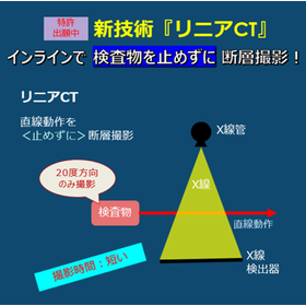
"Linear CT" is a new technology for tomography that performs inline inspection without stopping the inspection object.

Overview of new technology, benefits of implementation, and comparison images with conventional CT using battery cross-sectional images as an example!

We are currently developing a new technology called "Linear CT" (as of June 2025). In this article, we will provide an overview of this new technology, the benefits of its implementation, and comparison images with conventional CT using battery cross-sectional images as examples. If you are interested in "Linear CT," please feel free to contact us. *You can view the detailed content of the blog through the related links. For more information, please download the PDF or feel free to reach out to us.*

  • X-ray inspection equipment
  • X-ray Inspection Machine
  • CT scanner

Added to bookmarks

Bookmarks list

Bookmark has been removed

Bookmarks list

You can't add any more bookmarks

By registering as a member, you can increase the number of bookmarks you can save and organize them with labels.

Free membership registration

X-ray inspection Large X-ray CT system [Contracted inspection]

We provide X-ray inspection services! We have obtained certifications such as Nadcap and JISQ9100!

X-ray inspection is a non-destructive testing method that observes internal structures, casting and welding defects, and foreign objects from CT scan image data. The inspected object can be rotated 360 degrees, allowing for the acquisition of high-precision 3D images from multiple viewpoints. Equipped with one of the largest outputs in the country and high-resolution sensors, it can capture a variety of products. The maximum scan size is φ900mm × 1000mm, accommodating large products as well. Comprehensive aftercare is also provided by specialized inspectors.

  • Contract Inspection
  • CT scanner

Added to bookmarks

Bookmarks list

Bookmark has been removed

Bookmarks list

You can't add any more bookmarks

By registering as a member, you can increase the number of bookmarks you can save and organize them with labels.

Free membership registration

X-ray examination X-ray CT device [Contracted inspection]

We offer X-ray inspection services! We have obtained certifications such as Nadcap and JISQ9100!

The CT.Compact X-ray CT device is a radiation inspection technology that can obtain high-precision images while being compact and lightweight. Our company conducts contract inspections using this device, which is utilized in various fields such as manufacturing and the medical industry. The CT.Compact X-ray CT device can capture images at high speed while rotating the inspected object 360 degrees, allowing for non-destructive inspection of the internal structure, fine defects, and foreign objects of the inspected item. It also employs a highly sensitive image detector, enabling the acquisition of high-quality images. By utilizing advanced image processing technology, the brightness and contrast of the images can be freely adjusted. Our CT.Compact X-ray CT device is used for fine inspections of automotive parts, mechanical components, electronic parts, and more, thanks to its compact size and high-precision image acquisition capabilities. With advanced analytical technology and high-quality image acquisition capabilities, we can provide accurate and rapid inspections tailored to our customers' needs. We encourage you to make use of our CT.Compact X-ray CT device.

  • Contract Inspection
  • CT scanner

Added to bookmarks

Bookmarks list

Bookmark has been removed

Bookmarks list

You can't add any more bookmarks

By registering as a member, you can increase the number of bookmarks you can save and organize them with labels.

Free membership registration

Germany Large Linear Accelerator High Power Industrial CT diondo d7

High-performance linear accelerator CT device for the analysis and inspection of high-density components.

The Diondo D7 is developed for the analysis of inspection objects that are difficult to penetrate, particularly large specimens, and is suitable for analyzing high-density material components such as titanium, copper, steel, or thick aluminum (e.g., engines, crankcases, stators, turbine blades). The applications of this system primarily focus on component analysis to detect manufacturing defects and material flaws, as well as inspecting the shape and thickness of test specimens. Furthermore, the Diondo D7 can be equipped with linear accelerators of various performance levels to meet diverse customer needs. ▪ Linear Accelerator Performance levels of 3MeV, 6MeV, 9MeV, continuously adjustable ▪ Mechanical System Various sizes can be customized according to customer needs. ▪ Flat Panel Detector High-resolution (140μm) flat panel detector optimized for linear accelerator CT ▪ Line Array Detector High-resolution (200μm) linear array detector optimized for linear accelerator CT

  • Radiation detector
  • X-ray inspection equipment
  • Contract Analysis
  • CT scanner

Added to bookmarks

Bookmarks list

Bookmark has been removed

Bookmarks list

You can't add any more bookmarks

By registering as a member, you can increase the number of bookmarks you can save and organize them with labels.

Free membership registration

Observation of the internal state of delayed-release OD tablets.

The cause has become visually clear, contributing to the formulation of a policy for improvement at the customer level!

We will introduce a case that contributed to customer process improvement by capturing the changes in the internal structure of OD tablets before and after a rigorous test. X-ray CT is a non-destructive method that visualizes the spatial distribution of X-ray absorption rates within a sample. When changes occur inside the sample, there may be corresponding changes in the distribution of X-ray absorption rates, and by quantifying these changes, it is possible to infer what kind of changes have taken place inside the sample. The value "skewness," which represents the shape of the histogram of X-ray absorption rates within a region of a certain size, is one of the useful quantities that indicates the state of the sample's interior. 【Flow of Data Acquisition and Analysis】 ■Sample ■X-ray CT Observation ■3D X-ray Absorption Rate Data ■Data Analysis ■3D Distribution of Skewness of Absorption Rate Distribution in Multiple Regions *For more details, please refer to the PDF document or feel free to contact us.

  • X-ray Inspection Machine
  • CT scanner

Added to bookmarks

Bookmarks list

Bookmark has been removed

Bookmarks list

You can't add any more bookmarks

By registering as a member, you can increase the number of bookmarks you can save and organize them with labels.

Free membership registration

Investigation of Internal Defects in Metal Parts

You can check for the presence of internal defects without damaging the sample!

Regarding aluminum die-cast parts, information such as the presence of internal defects can be obtained through non-destructive observation using a 3D X-ray microscope (X-ray CT). Additionally, if defects are confirmed internally, the defects can be exposed and assessed through cross-sectional processing using techniques such as ion milling. When inclusions are present, the cause of occurrence can be estimated by confirming their composition. Please feel free to contact us if you have any inquiries. [Overview] ■ Acquisition of internal information through non-destructive observation (X-ray CT) ■ Cross-sectional structural analysis (cross-sectional processing + elemental analysis) *For more details, please download the PDF or feel free to contact us.

  • Other analysis and evaluation services
  • CT scanner

Added to bookmarks

Bookmarks list

Bookmark has been removed

Bookmarks list

You can't add any more bookmarks

By registering as a member, you can increase the number of bookmarks you can save and organize them with labels.

Free membership registration

パルスECT装置 Lyft

パルス ECT(PEC)の革新!CUIの測定が今、見直される

パルスECT装置Lyftは、パルスECT(PEC)技術を用いて、保温材下の母材(炭素鋼配管)の腐食を、保温材を取り外すことなくスクリーニング検査できる装置です。ポータブルタイプの世界最高水準・最新式機器であり、Grid mapping mode(分割計測)もしくはDynamic mode(連続計測)によるリアルタイムのC-スキャン画像化、高速のデータ取得(最大15points/s)を実現します。

  • Defect Inspection Equipment
  • CT scanner

Added to bookmarks

Bookmarks list

Bookmark has been removed

Bookmarks list

You can't add any more bookmarks

By registering as a member, you can increase the number of bookmarks you can save and organize them with labels.

Free membership registration

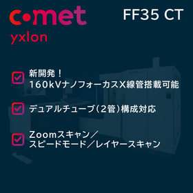
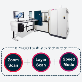
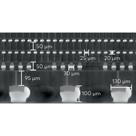
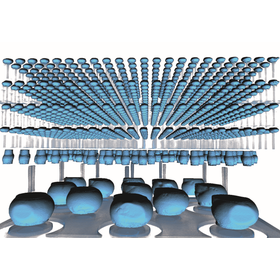
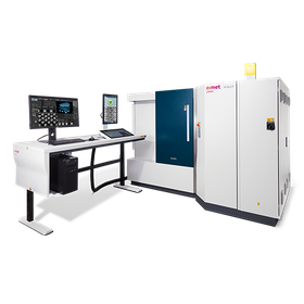
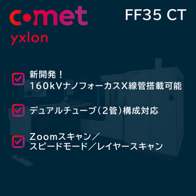
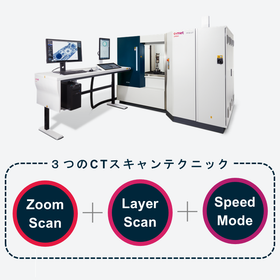
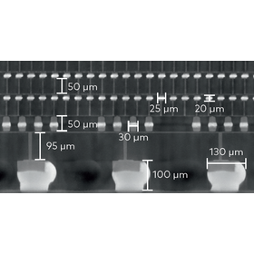
[For Cultural Heritage Survey] FF35 CT

Equipped with two types of X-ray tubes to visualize the internal structure of all cultural properties.

In archaeological cultural property surveys, it is essential to analyze the internal structure of valuable artifacts in detail and non-destructively. Understanding internal damage, manufacturing techniques, and past restoration history without compromising the value of cultural properties is crucial for their protection and research. The "FF35 CT" is equipped with two types of X-ray tubes, catering to diverse needs, and contributes to the detailed visualization of the internal structure of cultural properties, helping to solve these challenges. 【Usage Scenarios】 ■ Investigation of the internal structure of pottery and metal artifacts ■ Non-destructive analysis of mummies and wooden items ■ Identification of areas requiring restoration 【Benefits of Implementation】 ■ Visualization of internal structures without damaging cultural properties ■ Accurate assessment of deterioration and the need for restoration ■ Increased efficiency and quality of research

  • FF35CT?.png
  • FF35CT_3つのスキャン.png
  • スクリーンショット 2025-11-21 190949.png
  • スクリーンショット 2025-11-21 192111.png
  • FF35CT_SEMI_Product-Shot_large_500W.png
  • X-ray inspection equipment
  • CT scanner

Added to bookmarks

Bookmarks list

Bookmark has been removed

Bookmarks list

You can't add any more bookmarks

By registering as a member, you can increase the number of bookmarks you can save and organize them with labels.

Free membership registration

[For Research Institutions] FF35 CT: Improvement of Material Analysis Accuracy

FF35 CT with two types of X-ray tubes, catering to a wide range of material analysis needs.

In material analysis at research institutions, it is important to observe the internal structure of materials in detail and understand their properties. Particularly in the development of new materials and the performance evaluation of existing materials, technologies that can visualize internal structures non-destructively are required. The FF35 CT, with its dual tube configuration of 160 kV nano-focus and 225 kV micro-focus, covers a wide range from ultra-high resolution to thick samples with just one device. By observing the internal structure in detail non-destructively, it allows for the identification of failure causes before proceeding to destructive analysis, thereby improving the speed and success rate of the analysis. 【Usage Scenarios】 ■ Evaluation of internal structures in the research and development of new materials ■ Analysis of degradation or damage in existing materials ■ Detection of foreign matter contamination and defects 【Benefits of Implementation】 ■ Visualization of fine structures within materials enables detailed analysis ■ Non-destructive testing allows for the evaluation of valuable samples as they are ■ Improved research efficiency through early detection of failure causes

  • FF35CT?.png
  • FF35CT_3つのスキャン.png
  • スクリーンショット 2025-11-21 190949.png
  • スクリーンショット 2025-11-21 192111.png
  • FF35CT_SEMI_Product-Shot_large_500W.png
  • X-ray inspection equipment
  • CT scanner

Added to bookmarks

Bookmarks list

Bookmark has been removed

Bookmarks list

You can't add any more bookmarks

By registering as a member, you can increase the number of bookmarks you can save and organize them with labels.

Free membership registration