We have compiled a list of manufacturers, distributors, product information, reference prices, and rankings for Imaging.
ipros is IPROS GMS IPROS One of the largest technical database sites in Japan that collects information on.

Imaging Product List and Ranking from 12 Manufacturers, Suppliers and Companies

Last Updated: Aggregation Period:Sep 17, 2025~Oct 14, 2025
This ranking is based on the number of page views on our site.

Imaging Manufacturer, Suppliers and Company Rankings

Last Updated: Aggregation Period:Sep 17, 2025~Oct 14, 2025
This ranking is based on the number of page views on our site.

  1. null/null
  2. 一般財団法人材料科学技術振興財団 MST Tokyo//Testing, Analysis and Measurement
  3. ショーシンEM Aichi//others
  4. 4 日本レーザー Tokyo//Electronic Components and Semiconductors
  5. 5 日本ウォーターズ(米国本社 Waters Corporation)  Tokyo//Testing, Analysis and Measurement

Imaging Product ranking

Last Updated: Aggregation Period:Sep 17, 2025~Oct 14, 2025
This ranking is based on the number of page views on our site.

  1. Easy live cell imaging using an all-in-one fluorescence microscope.
  2. Live cell imaging CytoSMART Multi Lux2 ショーシンEM
  3. [Analysis Case] Evaluation of Composition and Impurity Distribution of ZnO Films by SIMS 一般財団法人材料科学技術振興財団 MST
  4. 4 Large-diameter imaging SIMS using RAE detectors 一般財団法人材料科学技術振興財団 MST
  5. 4 ImageXpress Micro4 High Content Imaging モレキュラーデバイスジャパン 東京本社

Imaging Product List

1~15 item / All 21 items

Displayed results

Life Science Solutions: Microscopy and Imaging

Excite the fluorophore at an optimal wavelength! Combine multiple plug-and-play wavelengths.

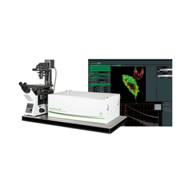
We would like to introduce our "Microscopy and Imaging." Achieve tighter focus and clearer images by exciting fluorophores at optimal wavelengths. Choose from a full range of scanning and imaging lasers for various types of confocal microscopes, not only for super-resolution methods like STED and PALM/STORM. 【Features】 ■ Excellent beam quality ■ More options ■ Fiber output ■ From ultraviolet to infrared *For more details, please refer to the PDF document or feel free to contact us.

  • solutions-life-sciences-microscopy-and-imaging-ultraviolet-to-infrared.jpg
  • 1.png
  • image_12.png
  • image_13.png
  • Laser microscope

Added to bookmarks

Bookmarks list

Bookmark has been removed

Bookmarks list

You can't add any more bookmarks

By registering as a member, you can increase the number of bookmarks you can save and organize them with labels.

Free membership registration

Life Science Solutions: Microscopy and Imaging

Combine multiple plug-and-play wavelengths! Achieve tighter focus and clearer images.

We offer not only super-resolution methods such as STED and PALM/STORM but also scanning and imaging lasers for various types of confocal microscopes. With excellent beam quality, you can achieve tighter focus and clearer images. Featuring fiber output, it combines multiple plug-and-play wavelengths. 【Features】 ■ Excellent beam quality ■ Fiber output ■ From ultraviolet to infrared ■ Fiber cable connection ■ Wavelength combinations *For more details, please refer to the related links or feel free to contact us.

  • Laser Components

Added to bookmarks

Bookmarks list

Bookmark has been removed

Bookmarks list

You can't add any more bookmarks

By registering as a member, you can increase the number of bookmarks you can save and organize them with labels.

Free membership registration

[Analysis Case] Three-Dimensional Distribution Evaluation of Dopants in MEMS Using SIMS

Imaging SIMS allows for the visualization of concentration distributions of trace elements in micro-regions.

We conducted three-dimensional imaging SIMS measurements of commercial MEMS products for B and As (measurement area: 75μm square, depth: approximately 1.5μm). After data processing, it is possible to extract surface distributions at arbitrary cross-sections and depths, depth direction distributions in arbitrary areas, and line profiles at arbitrary locations. Note: Since the sample is being excavated with an ion beam while capturing images in the depth direction, this constitutes destructive analysis.

  • Contract Analysis

Added to bookmarks

Bookmarks list

Bookmark has been removed

Bookmarks list

You can't add any more bookmarks

By registering as a member, you can increase the number of bookmarks you can save and organize them with labels.

Free membership registration

[Analysis Case] Dopant Investigation in NPT-IGBT Using SIMS

Evaluation of localized elements is possible with imaging SIMS.

Imaging SIMS measurements were conducted on a 50μm square area on the emitter side of the NPT-IGBT. Figure 1 shows the ion images of 11B and As obtained from the analysis. It can be seen that 11B and As are injected into the same area. Additionally, while conventional analysis calculates the average concentration of each element over the entire detection area, imaging SIMS measurements allow for the extraction of partial depth profiles, enabling the evaluation of the concentration distribution of dopants localized in the plane (Figure 2).

  • Contract Analysis

Added to bookmarks

Bookmarks list

Bookmark has been removed

Bookmarks list

You can't add any more bookmarks

By registering as a member, you can increase the number of bookmarks you can save and organize them with labels.

Free membership registration

Large-diameter imaging SIMS using RAE detectors

SIMS: Secondary Ion Mass Spectrometry

Imaging SIMS is effective for obtaining spatial distribution information of hydrogen and impurities at ppm levels. By using a RAE (Resistive Anode Encoder) detector for projection-type imaging SIMS, it is possible to obtain distribution images with a larger diameter and deeper regions compared to the commonly used scanning methods in imaging analysis.

  • Contract Analysis

Added to bookmarks

Bookmarks list

Bookmark has been removed

Bookmarks list

You can't add any more bookmarks

By registering as a member, you can increase the number of bookmarks you can save and organize them with labels.

Free membership registration

ImageXpress Micro4 High Content Imaging

Supports a wide range of applications! Enables high-speed image acquisition.

This product is a system that has perfected image acquisition and analysis technology. Thanks to its agile design, high-speed image acquisition has become possible. It comes with an option to upgrade to a confocal system as needed. 【Features】 ■ Beautiful image acquisition and expandability with the addition of a confocal unit ■ Compatible with a wide range of applications through objective lenses from 1x to 100x and a customizable 5-position filter cube ■ Fast data acquisition with a wide field of view and a broad dynamic range ■ Expand applications by adding a bright field unit, liquid dispensing mechanism, and environmental control unit *For more details, please refer to the PDF document or feel free to contact us.

  • Image Cytometer

Added to bookmarks

Bookmarks list

Bookmark has been removed

Bookmarks list

You can't add any more bookmarks

By registering as a member, you can increase the number of bookmarks you can save and organize them with labels.

Free membership registration

Vintasoft

Developing professional image acquisition for the web!

Vintasoft develops professional Image Acquisition, Barcode Reader and Generator, and Document Imaging SDKs for .NET, WPF, and WEB. We offer products such as the impressive and user-friendly "Vintasoft Imaging .NET SDK" and "Vintasoft Barcode .NET SDK." Please feel free to contact us if you have any inquiries. 【Features】 ■ Type: Shareware ■ Language: English ■ Operating Environment: Windows, Linux, macOS *For more details, please refer to the related links or feel free to contact us.

  • Company:B7
  • Price:Other
  • Software (middle, driver, security, etc.)

Added to bookmarks

Bookmarks list

Bookmark has been removed

Bookmarks list

You can't add any more bookmarks

By registering as a member, you can increase the number of bookmarks you can save and organize them with labels.

Free membership registration

Technical Team "morpho"

Introduction of automatic checks using AI eyes in video surveillance operations! Improving productivity and addressing the shortage of successors in technology.

"morpho" is a team of engineers in the field of "Imaging AI," which combines digital image processing technology with advanced artificial intelligence (AI) and deep learning. By using AI to automatically classify inspection images, it becomes possible to accelerate and enhance the precision of the inspection process, achieving an increase in the number of inspections and a reduction in the duration simultaneously. In jobs that require skilled techniques, AI support allows even those with less experience to handle tasks, leading to improved productivity and addressing the shortage of successors in technology. 【Features】 ■ Enables acceleration and high precision of the inspection process ■ Achieves an increase in the number of inspections and a reduction in duration simultaneously ■ Improves the accuracy of routine tasks and enhances the working environment ■ Allows for task handling even with less experience ■ Contributes to increased productivity and addresses the shortage of successors in technology *For more details, please download the PDF or feel free to contact us.

  • 16-2.PNG
  • others

Added to bookmarks

Bookmarks list

Bookmark has been removed

Bookmarks list

You can't add any more bookmarks

By registering as a member, you can increase the number of bookmarks you can save and organize them with labels.

Free membership registration

Fluorescence microscopy: single-molecule microscopy, time-resolved, super-resolution imaging.

The fluorescence microscope "MicroTime series" offers a variety of solutions for time-resolved confocal microscopy.

MicroTime Series A high-sensitivity microscope for single molecules equipped with picosecond time resolution and super-resolution imaging capabilities. ■ MicroTime 200 (Single Molecule Sensitivity Complete Time-Resolved Confocal Fluorescence Microscope) ◎ Complete system with laser coupling unit ◎ Multi-channel detection unit ◎ Single molecule sensitivity for excitation light from 266 to 900 nm ■ MicroTime 200 STED (Super-Resolution Time-Resolved Confocal Fluorescence Microscope) ◎ Confocal STED system ◎ Optical resolution of less than 50 nm ◎ Supports gate STED (gSTED) and gSTED-FCS ■ MicroTime 100 Upright Time-Resolved Fluorescence Microscope ◎ Laser coupling unit / Multi-detector option capable of up to 4-channel detection ◎ Pulsed diode lasers (wavelength: 375 nm to 810 nm) ◎ XY scanning piezo stage for 2D lifetime imaging ■ Sympo64 Fluorescence Lifetime Software and Correlation Software available * Upgrade kits for laser scanning microscopes from major manufacturers are also available * A PDF catalog can be downloaded; for details, please contact us.

  • Laser microscope

Added to bookmarks

Bookmarks list

Bookmark has been removed

Bookmarks list

You can't add any more bookmarks

By registering as a member, you can increase the number of bookmarks you can save and organize them with labels.

Free membership registration

Synthetic Technology: Molecular Imaging

We will strongly support research and development in the field of molecular imaging!

Molecular imaging refers to a technique that non-invasively visualizes changes in molecular groups within living organisms (such as enzymes, genes, membrane proteins, etc.) and observes them dynamically and quantitatively. A representative method in this field is positron emission tomography (PET), which is well-known in current research. In recent years, technologies utilizing fluorescence and near-infrared light have also been applied in the fields of biochemistry and regenerative medicine. As it lies at the intersection of medicine, pharmacy, and engineering, this field is expected to see further development. Nard Institute will continue to support this area from the standpoint of organic compound development. [Theme] ■ VARGULA LUCIFERIN ■ Fluorescent labeling of organelles *For more details, please refer to the related links page or feel free to contact us.

  • others
  • Other contract services

Added to bookmarks

Bookmarks list

Bookmark has been removed

Bookmarks list

You can't add any more bookmarks

By registering as a member, you can increase the number of bookmarks you can save and organize them with labels.

Free membership registration

Target mass spectrometry imaging (MSI)

Utilization of overwhelming reliability through overwhelming sensitivity and speed - Target MS imaging capable of quantification.

Mass spectrometry imaging (MSI) has often utilized MALDI as an ionization method, but it requires specialized knowledge and expertise for matrix selection and application. On the other hand, DESI applies ESI technology, eliminating the need for a matrix and enabling the ionization of a wide range of molecules. DESI XS MSI has been applied in combination with time-of-flight (ToF) mass spectrometers to directly map molecules in tissue sections, making it ideal for non-target exploratory analysis. With the recent possibility of combining it with tandem quadrupole (TQ) mass spectrometers, the sensitivity, speed, and quantitativeness of target molecule measurements using multiple reaction monitoring (MRM) mode have improved, allowing for widespread application in various fields such as drug analysis, biomarker detection, and surface analysis of materials, focusing on the "localization + quantification" of molecules. A remarkable and unique method has become available to quantitatively clarify the localization of previously unseen molecules. 【Features】 - High-sensitivity, rapid, and highly selective visualization of molecular localization - Improved sensitivity and spatial resolution with a new high-performance sprayer - No complicated sample pretreatment required, enabling comprehensive and detailed spatial information acquisition

  • Analytical Equipment and Devices

Added to bookmarks

Bookmarks list

Bookmark has been removed

Bookmarks list

You can't add any more bookmarks

By registering as a member, you can increase the number of bookmarks you can save and organize them with labels.

Free membership registration

[Analysis Case] Evaluation of the BSF Layer in Si-based Solar Cells

Impurity evaluation with in-plane distribution is possible using cross-sectional imaging SIMS.

This paper introduces a method for evaluating the in-plane distribution of solar cells with surface roughness while avoiding the effects of the roughness, using cross-sectional samples assessed by imaging SIMS. It was found that in regions identified as p+ Si layers by cross-sectional SCM, B and Al were present in high concentrations. Furthermore, using line profiles, the dopant distribution was converted to concentration, quantifying the degree of inhomogeneity in the plane. This method can also be applied to deep junction evaluations of power devices and impurity distribution assessments between and within trenches. Measurement methods: SIMS, SCM, SNDM Product field: Solar cells Analysis objectives: Trace concentration evaluation, composition distribution evaluation, shape evaluation For more details, please download the materials or contact us.

  • Contract Analysis

Added to bookmarks

Bookmarks list

Bookmark has been removed

Bookmarks list

You can't add any more bookmarks

By registering as a member, you can increase the number of bookmarks you can save and organize them with labels.

Free membership registration

[Analysis Case] Evaluation of Composition and Impurity Distribution of ZnO Films by SIMS

Visualization of in-plane distribution through imaging SIMS analysis.

The uniformity of the film composition and the distribution of impurities, which are one of the elements in device creation, were evaluated using imaging SIMS analysis. Through data processing after measurement, we can obtain planar images (Figure 1), cross-sectional images (Figure 2), depth distribution profiles at arbitrary locations (Figures 3 and 4), and line profiles. From the distribution of constituent materials and impurities, we can gain information that leads to process and film quality improvements.

  • Contract Analysis

Added to bookmarks

Bookmarks list

Bookmark has been removed

Bookmarks list

You can't add any more bookmarks

By registering as a member, you can increase the number of bookmarks you can save and organize them with labels.

Free membership registration

[Analysis Case] Evaluation of SiC Power MOSFET Dopants by SIMS

Imaging SIMS enables the evaluation of localized elements.

We disassembled a commercially available SiC power MOSFET and conducted imaging SIMS measurements to evaluate the concentration distribution of the dopant elements Al, N, and P in a 20 µm square area to a depth of 0.5 µm. We will present a case where we extracted the depth profile concentration distribution of Al, N, and P localized within the sample surface from the data processing after the imaging SIMS measurements.

  • Contract Analysis

Added to bookmarks

Bookmarks list

Bookmark has been removed

Bookmarks list

You can't add any more bookmarks

By registering as a member, you can increase the number of bookmarks you can save and organize them with labels.

Free membership registration