Z Series Modular Zoom Stereo Microscope
All products are sold individually as standard specification items. We offer quick delivery due to complete stock availability.
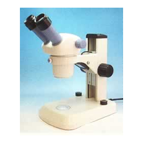
The Z series modular zoom stereo microscope is an ideal choice for building a new inspection system using user-friendly components. With three types of zoom range options and a combination of various objective lenses and eyepieces, the system can be customized to meet the true needs of the user. The optional photo port insert and C-mount adapter enable imaging with C-mount cameras. Additionally, external light sources such as section LED ring lighting and the same lighting equipped with polarizers/analysers are also available. For the product lineup, specifications, and sales prices of standard products, please visit the relevant product page on the official website of Edmund Optics Japan Co., Ltd. (accessible from the product homepage linked above). *You can download the catalog featuring this product from the link below for Edmund Optics Japan.
- Company:エドモンド・オプティクス・ジャパン
- Price:Other